XLMTM mechanism of disease

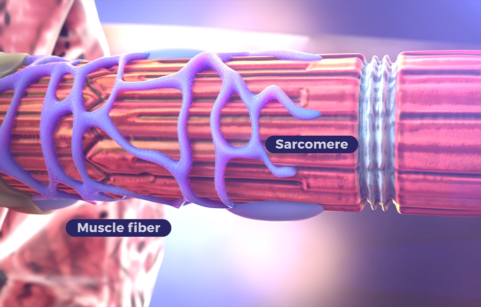
Skeletal muscle structure and function are affected by dysfunctional or absent myotubularin protein

Dysfunctional or absent myotubularin protein results in disorganized and defective muscle fibers. This leads to profound muscle weakness and hypotonia, impacting respiratory and neuromuscular motor function.1-4 Myotubularin is a phosphatase involved in several cellular processes required for skeletal muscle maintenance and function, including5-9:

  • Muscle growth and differentiation
  • Cellular organization and structure
  • Cellular function

Normal neuromuscular junction

Healthy

In healthy muscle cells, myotubularin protein associates with the muscle triad, which is a structure composed of Tatubules and the terminal sarcoplasmic reticulum.3 The triad mediates excitation-contraction (EC) coupling, a process in which electrical stimuli at the neuromuscular junction (NMJ) are translated into muscle contractions.5

Abnormal

In humans and animal models, defective myotubularin protein in skeletal muscle cells results in impaired EC coupling and mislocalization of cellular organelles. Skeletal muscle impairment, specifically EC coupling, is believed to be a primary cause of profound muscle weakness associated with X-linked myotubular myopathy (XLMTM).4

Understanding XLMTM

Learn about the underlying cause of disease and associated complications.

XLMTM pathology has the potential to be improved

Unlike other neuromuscular disorders, such as Duchenne muscular dystrophy (DMD) or spinal muscular atrophy (SMA), muscle fiber necrosis leading to atrophy is usually absent in XLMTM.10-12 Because the muscle fibers have not atrophied, the skeletal muscle defect in XLMTM is unique in its capacity to be improved by repairing function of the triad and recovering contractile force.3,13-16 Future therapies can potentially improve the pathology of XLMTM.

XLMTM presents with a range of signs and symptoms and has a substantial impact on the lives of patients and their families.

References

1. Al-Qusairi L, et al. Proc Natl Acad Sci U S A. 2009;106(44):18763-18768. 2. Dowling JJ, et al. PLoS Genet. 2009;5(2):e1000372. 3. Buj-Bello A, et al. Hum Mol Genet. 2008;17(14):2132-2143. 4. Zanoteli E. Expert Opin Orphan Drugs. 2018;6(6):375-384. 5. Dowling JJ, et al. In: Adam MP, et al, eds. GeneReviews®. 2002. 6. Buj-Bello A, et al. Proc Natl Acad Sci U S A. 2002;99(23):15060-15065. 7. Di Paolo G, et al. Nature. 2006;443:651-657. 8. Hnia K, et al. J Clin Invest. 2011;121(1):70-85. 9. Amoasii L, et al. J Cell Sci. 2013;126(Pt 8):1806-1819. 10. Lawlor MW, et al. J Neuropathol Exp Neurol. 2016;75(2):102-110. 11. North KN, et al. Neuromuscul Disord. 2014;24(2):97-116. 12. Guan X, et al. Methods. 2016;99:91-98. 13. Mack DL, et al. Mol Ther. 2017;25(4):839-854. 14. Maani N, et al. Nat Commun. 2018;9(1):4849. 15. Childers MK, et al. Sci Transl Med. 2014;6(220):220ra10. 16. Elverman M, et al. Muscle Nerve. 2017;56(5):943-953.

STAY INFORMED

Connect with us to get the latest information

Email Address*
Phone Number
Institution / Affiliation*

*Required fields

By submitting this form, you agree to allow Astellas Gene Therapies and its agents to collect the information provided and to be contacted directly by an Astellas Gene Therapies representative. Your information will not be used for any other purpose than for a representative to respond to your information request and/or to send you XLMTM updates if you have registered to receive them.

Astellas Gene Therapies will not sell, rent, or otherwise distribute your name and any personally identifiable information outside of Astellas Gene Therapies and its agents. Astellas Gene Therapies will only use your information in accordance with its Privacy Statement.